What is sciatica and what can I do at home to help?
- carmenmakepeace

- Apr 7, 2024
- 13 min read

Let's start with a definition.
Sciatica is a nerve like pain that radiates from the sciatic nerve typically from the buttocks region down the back of the upper thigh, referring further into the lower leg, calf and foot.
What's nerve pain feel like?

Nerve pain is often described as:
Pins & Needles in the limb
Sharp & Stabbing
Throbbing/Heated/Stabbing
Tingling in the skin
A Bee sting that travels along a limb.
Reduced sensation - the skin and limb feel "odd"
Lack of strength moving in a direction - this needs assessing
Numbness
Deadness to the area when you yourself touch it.
Burning pain (either icy or hot)
When I hear these words when a client is telling me about their experience my mind as a therapist says - ah nerve pain. Right let's think what could be the cause of this.
Whenever we think about nerve pain as a therapist we have to think about the nerve root and pathway through the tissues and all the structures surrounding it that can compress and restrict that nerve.
how do I know I have sciatica?

That's a really good question, because sciatica is compression & inflammation of the sciatic nerve that causes nerve pain down the back of the leg and predominately but not limited to the upper thigh.
It shouldn't be confused with any other nerve pain in the leg.
For instance, we can get nerve pain:
On the outside of the upper thigh
(On the line of where your jean's seam would be)
In the front of the thigh where our quads are
In the inner thigh and groin area
These are all examples of other nerve compressions similar but different to the sciatic nerve.
Sciatica is compression of the sciatic nerve leading to the symptoms stated of burning, pins and needles, throbbing...
So where is the sciatic nerve and how does it get compressed in the first place?

In order to explain how the sciatic nerve gets compressed in the first place I have to show you the road map of where it starts, where it journeys through and where the sciatic nerve ends!
1. the sciatic nerve starts in the spine & exits the back of the pelvis
The sciatic nerve starts in the spine, when the nerves between the last two lumbar vertebrae L4 and L5 join into the first three nerves of the sacrum S1, S2, S3 forming what's called the sacral plexus.***
***
Any plexus in the body is a word used where a group of spinal nerves merge like a train hub of interconnecting tracks. It builds resiliency into our systems so that if one of those spinal nerves was damaged our leg could still move in the case of the sciatic nerve.

2. The sciatic nerve travels through the back of the gluteal muscles....

3. in particular the piriformis...

The piriformis is a deep muscle of the lower third of the gluteal region (buttocks).
In different populations it has been accessed how much anatomical variation exists between the sciatic nerve position and the piriformis muscle.

CPD: common peroneal nerve TN: tibial nerve. (Poutoglidou, et al., 2020)
A: The common sciatic nerve presentation: 64-99% of the population dependent on geographic demographics
The majority of the population with the sciatic nerve travelling below the piriformis muscle undivided.
B: The sciatic nerve splits early above the knee: CPN through muscle
the sciatic nerve spilts into the Common Peroneal Nerve and exits through the piriformis muscle whilst the other half of the tibial nerve exits below.
C: The sciatic nerve spilts early above the knee: CPN above muscle
The common peroneal nerve exits above the piriformis muscle and the tibial nerve below.
D: The sciatic nerve runs through the piriformis
The sciatic nerve exits through the piriformis muscle.
E: The sciatic nerve splits early: CPN above the piriformis
The common peroneal nerve exits above the piriformis muscle and the tibial nerve runs through the muscle.
F: The sciatic nerve passes above the piriformis muscle
The sciatic nerve remains undivided but exits above the piriformis muscle.
These findings are particularly important as a future explanation of how the piriformis can compress the sciatic nerve.
4. And deep 6 rotators at the base of the sit bones...

5. the sciatic nerve travels through the back of the thigh:

6. and splits at the popliteal fossa above the knee supplying the calf...

7. Finally the calcaneal & plantar nerves supply the foot.
why is knowing the structures that the nerve passes through important?
Because in clinic we consider all the various places that the nerve pathway could be compressed to cause irritation...
what are the structures that can compress the sciatic nerve?

disc herniation from L2, L3, L4, L5.
sacroiliac dysfunction (where the sciatic nerve exits the pelvis)
compression from the piriformis muscle
compression from the deep 6 muscles
FASCIAl RESTRICTIONS ACROSS THE PELVIS & lower limb from trauma
spinal stenosis
trigger point patterns:
The erector spinae muscles
The quadratus lumborum
The ilio psoas muscle
The hamstring muscle group
The piriformis
The glute minimus muscle (fake sciatica)

what do we do in clinic to help?
With everyone that walks in with any type of pain we take a thorough case history
The reason for this is that the case history tells us so much.
What we are looking for is how the symptoms started.

particularly:
was there was a specific event that started the pain in recent history.
A fall or injury event:
"I was playing football the other night when I was tackled and twisted my ankle..."
"I picked up a piece of furniture awkwardly and felt a sudden sharp pain...."
"I don't normally run when it's tipping it down but anyway I twisted really weirdly and my leg went out from under me..."
Then we will assess how recently this happened, are we talking years ago or the other week?
If we're describing something last week...
I would suspect a disc herniation and structural changes
things that would be picked up in an x-ray, mri, ultrasound etc
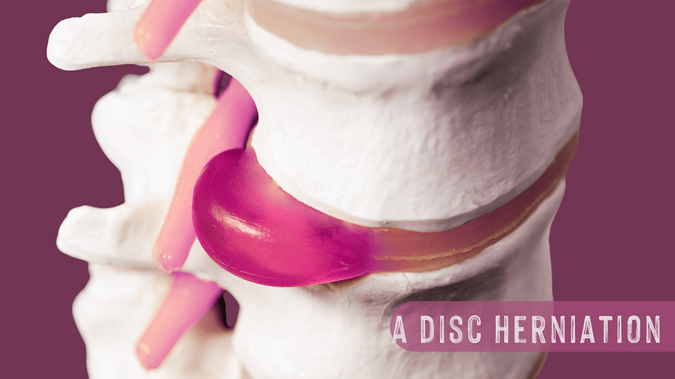
It's likely an acute injury has occurred with inflammation still occurring today and I am more likely to think of a disc herniation.
A disc herniation is where the inside of the disc (the nucleus pulposus) was compressed under so much load that it escaped it's confines of the disc's fibrous outer material and herniated outwards.
Typically this happens either to the left or the right of the spinal disc (It is very rare to occur to both sides at once).
In this instance immune cells arrive at the herniated material now outside the disc and start to clean up the debris.
This hardens that material and causes it to project onto the spinal cord compressing the nerves there.
Because the spinal cord actually terminates at the second lumbar vertebra (L2) in most adults and from there all the lower nerve fibers from L3 to S5 descend through the spine as separate nerves until their exit level...
Disc compression at L2 can actually cause compression and symptoms of sciatica, as can any disc compression lower down.
So in clinic we would consider all these spinal segments of the lower spine.
I would also check the Sacroiliac joints at the back of the pelvis

Sacroiliac joint dysfunction is where the two hip bones at the back of the pelvis are not functioning correctly with the bones at the base of the spine - the sacrum, they are either displaying too little motion or too much motion.
Classic signs of Sacroiliac joint dysfunction:
Pain standing for long periods of time,
Pain getting into and out of a car as you sit down and lift your leg in to join you (think getting into and out of a sports car being the worst!);
Pain turning over in bed at night down the leg.

Sacroiliac joint dysfunction can be acute or chronic and would depend on the case history.
If there was recent trauma to this site I would consider it acute and treat accordingly.
If there was trauma here years ago, falling off a horse and breaking a coccyx for example...
I would think that fascially there will be trauma embedded in the connective tissues creating strain and hardening in those tissues and disrupting the balance of the pelvis.

piriformis syndrome:
Occasionally overuse of the muscles in the glute region particularly the piriformis muscle that helps us rotate the leg outwards so the toes are pointing out with a straight leg can cause compression of the sciatic nerve.
This is more common in members of the population with variations of the piriformis anatomy as shown above.
These variations appears slightly more common in women however can happen in men as well.
A common sign of the piriformis muscle restriction is when a client has an extreme turn out in the leg when lying face up when relaxed. You often see this in ballet dancers.
I have also seen this in weightlifters who tend to rotate externally when pulling a heavy weight off the floor to create more stability in the hip.
I have seen this in runners who have retained this posture because of an older injury in a hip flexor or from a knee replacement.
I have seen a predisposition of external rotation in the leg after almost every hip replacement I've ever treated due to a thickening of scar tissue alongside greater tension in these muscles from the operation.
Finally I have seen this a lot post pregnancy in women because of changes in the pelvic floor muscles.

similar to piriformis syndrome: compression of the nerve by the deep 6 rotators.
These small but very important muscles help stabilise our pelvis and interconnect fascially with our pelvic floor.
During and after pregnancy back pain is often seen in clinic alongside sciatica symptoms and a hardening and constriction in these muscles on assessment.
FASCIAl RESTRICTIONS ACROSS THE PELVIS & lower limb from older trauma

This is where treatment gets much more complicated.
When the injury has just happened (in the acute phase) the body will go through a healing cycle initially of:
Hemostasis
Inflammation
Proliferation
Remodelling
Hemostasis:
The blood vessels in the area constrict to reduce further bleeding. Platelets from the blood then arrive at the site and alongside fibrin proteins coagulate to form a scab sealing the site from further damage and blood loss. This process can take up to 2 days.
Inflammation:
Blood vessels dilate to release white blood cells and other immune cells into the area to prevent any infections during the healing process. Redness, swelling and heat are usual symptoms of inflammation. The inflamatory phase can last between 6 days or more.
Proliferation:
This is a stage of new cells being laid down in the tissue structure that will one day become a scar particularly in any tissue other than skin, because the other tissues cannot replace themselves to the same extent.
Muscle cells in particular are replaced with collagen which creates fibrous scars that have a different and reduced elasticity than the surrounding muscle tissue. Proliferation usually takes 2 weeks or more after the inflammatory phase but is dependent on the trauma healing time.
Remodelling:
This stage can last for up to two or more years depending on the tissue healing time.
In this stage cells are being cleaned up that were excessively laid down and scar tissue is being fine tuned.

Treatment in the acute stage:
When the injury has just happened as long as we are outside of the inflammatory phase we can assist tissue healing in terms of direction under load and organisation.
Treatment can improve fluid flow in tissues that have had restriction or pressure from swelling thereaby reducing pressure on nerves that are compressed.
Treatment can assist in the tissue proliferation and remodelling phase by actively promoting correct laying down alignment and progressive load rehabilitation -optimising that muscle's strength in the range that it is meant to perform.
We can manage load such that you don't go back into an inflammatory response however if you do overdo it we can once more improve fluid flow and reduce swelling in the area.
In a chronic case ie when fascial restrictions have built up over time:

Treatment becomes a lot more complex, we have to assess how the strain pattern arrived in the body. How the force went in.
We have to assess how the body has adapted over time to the current and past demands placed on it and then from their form a picture of how the connective tissues are holding the body in support.
Fascial actively contracts when a trauma arrives both to prevent colossal injury but also to enable the body to keep moving somewhat afterwards and hold its structure.
This means if you did crack your coccyx years ago after falling off a horse, you might find that:
The sacrum (the part of the spine above the coccyx) has also been affected - shifting it minutely to the opposite side of where the force came in.
The scaro-iliac joint on the opposite side of where the force came in is locked.
The piriformis in that side of the gluteal muscles has tighted to reduce further movement.
The deep 6 have tightened to in relation to the piriformis
The pelvic floor which is fascially connected to the deep 6 has attapted
Affecting the sex organs and some of the lower abdominal organs.
The hip flexor (PSOAS) on the opposite side to the gluteal muscle adaptations is holding opposing tension to manage pelvic stability
As does the opposite quadratus lumborum muscles.
A thickening of the ligaments around the spine themselves can impinge on the spinal nerve roots exiting the spine thereby directly creating the effect alongside these other fascial chains.
The lower limb tissues may have adapted to the pelvic state increasing dynamic tension in the adductors (inner thight) of one leg and the hamstrings and lateral IT band of the other leg.
This list of adaptation could continue through the trunk and into the diaphragm and chest cavity.

When we treat we can activate the neurological contractile aspect of fascial tissues to take the leg into the directionality of the force that once came in and follow the spontaneous movement of the fascia into it's tissue memory.
We follow it all the way into the fascial twist until we are at the fulcrum of the point in which the original force is still being held in a equilibrium state.
In that place the body responds to start getting to work to release the tissue memory and with it the fascial strain.
spinal stenosis:

I placed spinal stenosis here because it would like the tension built in the fascial chain have occurred over a significant time frame and be chronic in nature.
Now it may have a prior history of trauma to the spine a long time ago in the past. (sometimes 20 plus years ago).
But as a form of spinal arthritis that causes a narrowing of the openings (foramina) of the spine that allow the nerve roots to pass out of the spine to form the sciatic nerve say, it can also compress that exiting nerve in a similar way as the disc herniation.
Spinal stenosis has symptoms of:
Pain in your lower back.
Pain that originates in your buttocks and extends down your leg towards your foot.
A heavy feeling in your legs, sometimes described as cramping.
Numbness or tingling (“pins and needles”) in the gluteals, leg or foot.
Pain that worsens on standing for a long duration, walking or walking downhill.
Pain that lessens when you lean forward, walk uphill or sit.
trigger point patterns:
The final option after all the above causes of nerve compression are trigger points in the soft tissues: muscles surrounding the sciatic nerve.
Of most clinic significance would be the piriformis, deep 6 rotators (which have the same referal pattern as the piriformis) and the hamstring muscles.
I have genuinely had a case with a client recently where they had spoken to a NHS physiotherapist on a zoom call and become so frustrated at a lack of clarity such that I had had the very worst ideas of structural deviations in my mind.
I went through all the options above and in the end treated trigger points in the soft tissues only to see the pain referral completely vanish by the end of the session.
It was a moment for me as a therapist to have confidence in the ability to know the structures involved but also to run through the list of options and check everything off systematically all whilst explaining to my client what I was doing and why.
Sometimes it can just be the simple option, which is always welcome, because it takes on average 1-3 sessions to completely resolve, whilst the other options can take longer.
The trigger point patterns below have the location of the muscle on the skeleton with the pain pattern next to them in red, showing the referral pattern that is most often felt.


what can we do at home to help ourselves?
In the most serious case - go to A&E:
Now I don't normally say this often but there is a big red flag here that needs to be clearly stated.
If you ever have the following symptoms in relation to this pain pattern down the back of the leg, it is not a time to see a manual therapist, you need medical intervention at Accident & Emergency.
If you have:
Nerve pain radiating down both legs at the same time (bi-lateral pain)
Numbness in your urinary and lower pelvis (especially when going to the toilet)
A lack or loss of bladder or bowel control (any amount of loss of control)
Unrelenting night pain (non-stop continuous extreme pain)
A suspected break and the complete inability to load bare on your legs.
Sudden unexpected weight loss (and please be clear you weren't on any form of calorie restriction).
Go to A&E
at A&E you will have a scan of the spine to assess whether cauda equina is present which requires medical intervention.

so if you don't have cauda equina what can we do in clinic?
Well one of the most important first steps is always trying to understand the problem.
If you have a scan result from A&E showing a disc herniation then you will know that it takes on average 2-12 weeks for your immune system to fully remove the protruding herniation. In that time we can do a lot to help.
1. we assess the cause together
If you don't come to your appointment with a scan we can run through all the options above together and narrow down the possible cause. You should leave with a sense of what the problem is and how to combat it from education.
2. we treat opening up tissues to enable you to access more movement
3. we teach spinal and joint opening exercises to facilitate more fluid flows to the area.
If you have arthritis around the spine and or fascial restrictions, the first thing we can do to combat it is very gentle spinal opening exercises and stretches at home in a pain free range without straining. This is because healthy joints have easy gliding movement. Hydration is also an important part of any fascial restriction and if you don't drink enough water, now is the time to start.
4. we teach fascial stretching that's specific to your postural patterns
If you have a twist of fascial restrictions from the lower leg up into the pelvis from past trauma then you and your therapist can cultivate a series of stretches that if repeated daily for 10 minutes could substantially retain your postural patterns that are specific to your needs.
5. we teach stretching specific to the piriformis and deep 6 rotators
If you have compression due to the piriformis and deep 6 rotators of the gluteal muscles then we can teach you stretches to eases these areas specifically.
6. we discuss mindfulness and meditation practices to combat trauma
If you have a history of trauma in the past, starting to participate in mindfulness exercises and meditation are a wonderful way to down regulate pain signals.
There are some fantastic resources online including the pain course available on the headspace app, additionally local mindfulness meditation, yoga and Qi gong are wonderful options too.
But more than anything learning to even take 3-10 deep breaths and stop whilst making your morning tea or coffee is genuinely a wonderful first step.


We hope this was helpful:
As ever this is a complex topic and we hope you've found this run through helpful to understand your experience.
There are multiple reasons why you are experiencing nerve pain in your leg and it's our job to help you find the answer.
Please keep an eye out as we expand these topics to the other nerve pathways that can be compressed in the leg.
As ever it is difficult to do this alone. I myself seek treatment and need help remaining objective with my own challenges, if you need help please don't hesitate to reach out.
More than anything if you don't have the answers yet, trust yourself to know that you are the expert in how your body feels and your experience is real.
Much love
Carmen

References: click to expand
Poutoglidou, F., Piagkou, M., Totlis, T., Tzika, M., & Natsis, K. (2020). Sciatic Nerve Variants and the Piriformis Muscle: A Systematic Review and Meta-Analysis. Cureus, 12(11), e11531. https://doi.org/10.7759/cureus.11531


Comments